This website uses cookies to ensure you get the best experience on our website. Learn more
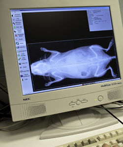
The assay is developed to provide high resolution images of the whole mouse skeleton. Analysis of the digital X-ray pictures is generally performed with respect to bones from the head (zygomatic bone, maxilla, mandibles), teeth, scapulae, clavicle, ribs (number, shape, fusion), pelvis, vertebrae (numbers, shape and potential fusion of cervical, thoracic, lumbar, pelvic and caudal ones), limb bones (humerus, radius, ulna, femur, tibia), joints, digits and syndactylism.
X-Ray MX-20 Specimen (Faxitron, Tucson, Arizona, USA)
At least 5 mice per group are required for appropriated analysis.
![]() Bone mineral density in female mice from different strains
Bone mineral density in female mice from different strains
Bone density scanning, also called dual-energy x-ray absorptiometry (DXA or DEXA) is used to measure bone mineral density. DEXA is also effective in tracking the effects of treatment for osteoporosis and other conditions that cause bone loss. The DEXA scan analysis can be also used to value the body composition in lean and fat tissue.
pDEXA Sabre (Norland)
Bone architecture may be analysed by both in vivo and in situ using Micron-scale X-ray Computed Tomography (also called µCT). It is particularly helpful for bone dynamics analysis. This augments our current capacities of skeletal examinations by TRAP histological stain, X-ray, DEXA scanning, quantitative NMR, and adapted clinical chemistry analysis.
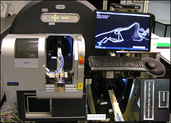
µCT (PerkinElmer Quantum FX, Waltham, Massachusetts)


Representative 3D volumes illustrating trabecular microarchitecture in the distal femur (left) and caudal vertebra (right) of Sham-operated and Ovarectomized (OVX) mice, 9 weeks after surgery. Similar volumes are used for morphometric quantification.

Example of longitudinal changes in trabecular bone volume fraction (BV/TV) and trabecular number (Tb.N) for Sham and OVX groups.

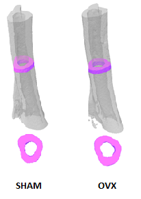
Representative 3D volumes illustrating cortical bone in the tibia of Sham-operated and Ovarectomized OVX mice, 9 weeks after surgery. The illustrations were generated by Alexandru Parlog (PHENOMIN-ICS).

Example of longitudinal changes in cortical bone volume fraction (BV/TV) and cortical thickness (Cr.Th) for Sham-operated and Ovarectomized OVX groups.
Contact us for help in designing your experiments on bone dynamics.
